Fluorescein Angiography for Diagnosis of Retinal Diseases
At Chugh Eye Surgery Centre, we offer Fluorescein Angiography (FA), a specialized imaging technique used to examine the circulation of the retina and choroid. It helps in diagnosing and managing various retinal conditions accurately and effectively.
What is Fluorescein Angiography?
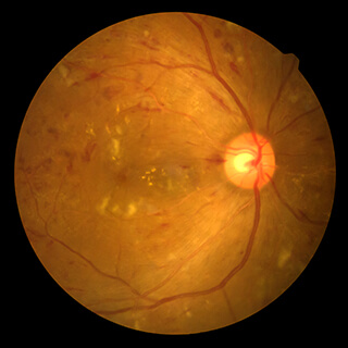
Fluorescein Angiography involves injecting a fluorescent dye into a vein in your arm. As the dye travels through the blood vessels in your eye, a special camera captures detailed images. These images help in identifying any blockages, leakages, or abnormal blood vessels in the retina and choroid.
When is Fluorescein Angiography Needed?
- Diabetic Retinopathy
- Age-Related Macular Degeneration (AMD)
- Retinal Vein Occlusions
- Macular Edema
- Unexplained Vision Loss
- Choroidal Neovascularization
Benefits of Fluorescein Angiography
- Accurate diagnosis of retinal disorders
- Guides treatment planning for retinal diseases
- Non-invasive and quick procedure
- Early detection of vision-threatening conditions
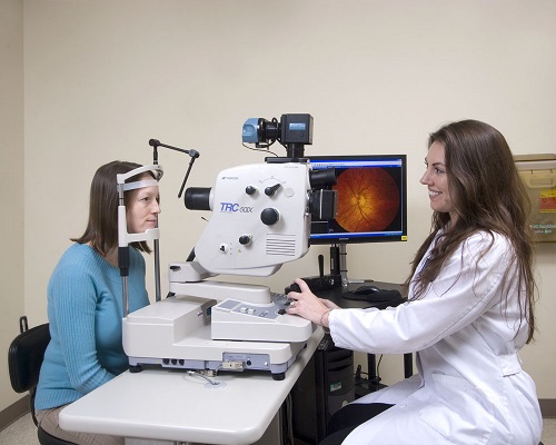
What to Expect During the Procedure?
- Fluorescein dye is injected into a vein in your arm.
- A special camera takes rapid pictures of the dye passing through the blood vessels in your eye.
- The procedure usually takes about 10-20 minutes.
- Temporary yellowing of the skin and urine discoloration may occur, which resolves quickly.
Post-Procedure Care
- You can resume normal activities immediately after the test.
- Drink plenty of water to help flush the dye from your system.
- If you experience any discomfort or unusual symptoms, inform your eye doctor.
Why Choose Chugh Eye Surgery Centre?
- Experienced retinal specialists
- Advanced imaging technology
- Comprehensive evaluation and diagnosis
- Patient-centered care with personalized attention